 African Spiny Mice: 'Making incremental - but essential - progress toward answering evolutionary questions about this extraordinary mammal' (Getty Images)
How does a five-inch long rodent native to African and Middle Eastern deserts offer what researchers affiliated with the Brotman Baty Institute believe is the greatest hope for restoring a person’s damaged or failing kidneys, liver, or other internal organs?
That is the question, as intriguing as it is complex, that Drs. Elizabeth Nguyen and Mark Majesky are determined to answer. In collaboration with colleagues from the University of Washington, Stanford University, the Howard Hughes Medical Institute, and Northern Arizona University, they recently published a preprint paper creating the highest quality reference genome on the African Spiny Mouse, the world’s only mammal able to naturally regenerate organs, as well as muscles, hair, spinal cord, and skin.
“This paper is just the beginning,” said Nguyen, an acting assistant professor of pediatric nephrology at UW Medicine and an investigator at Seattle Children’s. “Our work is opening doors for us – and others – to create new experiments as we understand more about this rodent and, in turn, develop potential targeted therapies for humans.”
BBI played an important role in opening those doors. Work on the paper was supported through the UW Oxford Nanopore Sequencing Core, which was created, in part, with BBI support and which provides a subsidy for investigators at UW Medicine, Seattle Children’s Research Institute, and the Fred Hutchinson Cancer Center. This subsidy offset some costs of sequencing conducted by the lab of BBI’s Dr. Danny Miller.
“This is the first time we've done Nanopore sequencing of this species,” Miller said. “I'm hoping we can do more. It's such an interesting animal with so much potential for helping us understand the response to tissue injury.”
That interesting species – with its large, pointy ears; curious dark eyes; and all but completely bald tail – date back somewhere between 5 and 11 million years ago. But it has been only 11 years since the scientific community discovered its unique biological function to regenerate skin. Researchers at the University of Florida in Gainesville published findings and observations in the journal Nature, concluding: “As re-emergent interest in regenerative medicine seeks to isolate molecular pathways controlling tissue regeneration in mammals, (this mouse) may prove useful in identifying mechanisms to promote regeneration in lieu of fibrosis and scarring.”
That first paper, Majesky said, “showed that spiny mice shed their dorsal skin to avoid predators and then regenerate that lost skin without scar formation.” Subsequent studies led to discoveries of the mouse’s ability to regenerate damaged organ structure and function, as well as its spinal cord. For Majesky, those discoveries were revelatory.
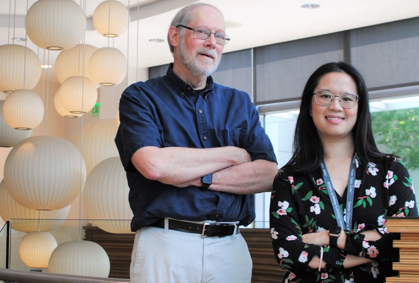
 Drs. Mark Majesky (left) and Elizabeth Nguyen: 'Restoration of solid organ architecture and function after injury or disease remains the holy grail of regenerative medicine.'
“Restoration of solid organ architecture and function after injury or disease remains the holy grail of regenerative medicine,” said Majesky, a professor of pediatrics and of laboratory medicine and pathology at UW Medicine. He is also the director of the Myocardial Regeneration Initiative and principal investigator in the Center for Developmental Biology & Regenerative Medicine at Seattle Children’s Research Institute.
The preprint paper, on which Nguyen and Miller are the lead authors, represents a significant step forward to finding that holy grail.
Long-read DNA sequencing was performed on blood samples in adult male spiny mice to generate the reference genome. In addition, to confirm the quality of assembly and annotation, as well as to identify a broad range of expressed transcripts, the team performed RNA sequencing of several tissues including brain, heart, liver and testis, to align to the genome.
“Hence, our highly contiguous and high-quality genome presented here will broadly benefit the growing (spiny mouse) community, and accelerate more detailed investigations into the genetic and epigenetic mechanisms underlying (the animal’s) novel capacity to maintain organ regeneration as adult mammals,” the authors wrote.
This is not the first time Nguyen and Majesky have collaborated on studies exploring the organ regeneration in the African Spiny Mouse. In November of 2021, working alongside Dr. Daryl Okamura, an assistant professor of pediatrics at the UW School of Medicine and an as an attending physician at Seattle Children’s, they published in iScience a paper demonstrating the rodent’s ability to “fully regenerate kidney structure and function,” noting its “genome appears poised at the time of kidney injury to initiate regeneration.”
So, what further analysis is needed to move the study of the African Spiny Mouse the next level leading to potential human trials?
Majesky outlines four important milestones:
- Identifying the critical sequences in the mouse’s genome that, collectively, initiate regeneration without fibrosis after acute organ injury
- Discerning the interplay between the DNA sequence itself and the epigenome that plays a critical role in which genes are turned on or off after tissues are damaged
- Determining the role and effect of myofibroblasts, the primary cell type responsible for enhanced collagen production in fibrosis, in the spiny mouse’s complex biological system of scar less wound healing
- Understanding how regenerating vascular cells and circulating immune cells, which both carry regenerating genomes, execute the complex interplay required for tissue regeneration.
“Everything about this animal is unique,” said Nguyen. “We are making incremental - but essential - progress toward answering evolutionary questions about this extraordinary mammal and using those answers to advance human regenerative medicine.”


