
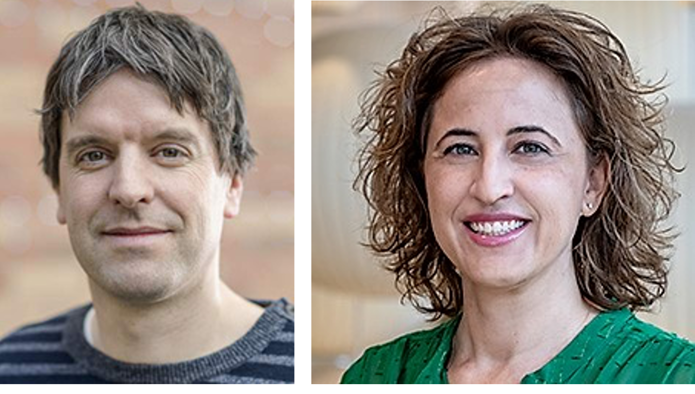
 BBI Members Drs. Kimberly Aldinger and Georg Seelig: Their first meeting was ‘a natural and serendipitous connection, a pivotal moment’ leading to an international research collaboration.
“By accessing fetal brain samples from both locations, here in Seattle and in the UK, we were able to go into great depth with our analyses of early cerebellum development.” --Dr. Kimberly Aldinger, Investigator, Seattle Children’s Hospital
A young child’s cerebellum is only one-fourth of its adult size, yet it contains the blueprint for integrating environmental cues with developing motor, cognitive, and emotional skills that influence the rest of that child’s life. That development begins 30 days after conception and continues through the child’s second birthday.
It is an extraordinary biological process that fascinates Dr. Kimberly Aldinger, an investigator at Seattle Children’s Hospital and member of the Brotman Baty Institute for Precision Medicine (BBI). She and other BBI members, as well as other researchers at Seattle Children’s, Yale University, and schools in the United Kingdom, published a paper last August in Nature Neuroscience after studying a 13-week window of fetal cerebellum development. Those other BBI members include: Drs. Dan Doherty, Ian Glass, and Kathleen J. Millen. In labs nearly halfway around the world from each other, Aldinger and colleagues mapped genes associated with neurodevelopmental and adult-onset neurodegenerative disorders to relevant cell types, as well as created a framework for verifying lineage relationships associated with brain abnormalities.
“By accessing fetal brain samples from both locations, here in Seattle and in the UK, we were able to go into great depth with our analyses of early cerebellum development,” said Aldinger. “This collaboration was not only unique and helpful, it also exemplified our international partnership.” Indeed, that partnership may be equally as interesting as the researchers’ findings. The catalyst for this four-year endeavor was a flyer Aldinger saw in November of 2017 announcing a presentation by Dr. Georg Seelig another BBI member at the University of Washington. She decided immediately to attend, thinking, “Oh, you have information on mouse cerebellum, and we’re looking at this in humans.”
Seelig, a professor with dual appointments at UW Department of Electrical & Computer Engineering and the Paul G. Allen School of Computer Science & Engineering, remembers the encounter. “Kim pointed that one of the slides in my presentation was incorrect because of an error in the specific location of a neuron,” Seelig said.
Aldinger recalls that the presentation – and her conversation with Seelig afterward – was “a natural and serendipitous connection – a pivotal moment.”
That initial connection led to a meeting with Aldinger and her colleagues and Seelig’s lab members to discuss the assay used in the mouse study. The meeting spawned the first of several experiments; she and a technical specialist brought samples of fetal cerebellums to the lab. Aldinger said it was questionable whether her fetal samples would perform as well as mouse tissue, noting the assay was not designed for human brain tissue.
“But because our samples were collected within two hours of the procedure, we had very high-quality samples,” she said. “That worked to our advantage. We were the first group outside of their lab to perform the assay to see how translatable it was.”
The project started using samples from the UW’s Birth Defects Research Laboratory (BDRL) collected over three years.
“We accessed the highly technical capabilities of BDRL in addition to its unique tissue procurement,” Aldinger said. “BDRL used laser capture microdissection to cut out specific regions of the cerebellum tissue, then we did bulk RNA-sequencing to determine the gene expression profiles associated with specific regions within the cerebellum.”
At the same time, Dr. Parthiv Haldipur, a neuroscientist at Seattle Children's Research Institute, had been working with colleagues at Newcastle University, a public research university based in Newcastle upon Tyne in Northeast England, and University College in London. Both schools hold a similar repository of fetal tissue, the Human Developmental Biology Resource.
By accessing tissues from both locations, Aldinger and her team obtained a sufficient number of tissues to study fetal cerebella in-depth, allowing them to have tissue samples ranging in age from nine to 10 weeks post-conception. Haldipur, an honorary research associate at University College in London, noted that the fetal cerebellum samples from the UK were comparable in quality to those of the UW.
“By providing us with tissue for analysis, our collaborators in the UK and the University of Washington offered a unique and highly valuable resource,” Haldipur said. “We have developed excellent working relationships with the coordinators at each site and over the years, maximized the use of preserved specimens. Through our collaboration with bioengineers at UW we were able to implement the latest techniques to process tissue and sequence RNA from single cells.”
Haldipur and Aldinger have “synergistic expertise and perspectives” regarding cerebellar development, Aldinger said. He is versed in anatomy and histopathology, while Aldinger’s expertise is in molecular genetics, especially transcriptomics. They approach their research, she said, in different, but complementary ways.
Moreover, Haldipur worked closely on this study with Dr. Paula Alexandre of the University College in London’s Institute of Child Health. Alexandre is devoted to understanding the cellular and molecular mechanisms underlying brain cells’ behavior, proliferation and differentiation. This project with Haldipur and Aldinger was an obvious complement to her research, and she sees the likelihood of working together in the future.
“We will make use of our complementary knowledge and skills and I expect we will continue working together on the characterization of human cerebellum development,” Alexandre said.
Haldipur put the study Aldinger led into context for researchers focusing on the first three years of the brain’s development.
“This paper provided valuable information on the changes in cellular diversity taking place during normal human cerebellar development,” he said. “If we are to understand the mechanisms of human disease, including devastating childhood tumors and cerebellar birth defects, it is important to extend our work beyond the characterization of normal development. This study is only the tip of the iceberg.”

